"Pneumothorax is colloquially known as a lung burst or lung deflation. "
Pneumothorax, colloquially known as a burst lung or collapsed lung, is a condition caused by tears in the lining of the lung or bubbles in the lung. When this happens, air accumulates in the lung and the lung becomes partially or completely deflated. Pneumothorax can occur spontaneously, traumatically, secondary to various diseases or as a result of medical interventions.
Symptoms of pneumothorax include shortness of breath, chest pain, difficulty breathing, coughing, weakness, fatigue and dizziness. These symptoms can range from mild to severe.
What are the Causes of Pneumothorax?
The causes are very diverse. In young patients, the cause is the bursting of small air bubbles called lung bullae and blebs, while in older patients it is usually COPD. Smoking is highly associated with the disease and is effective in the growth and subsequent rupture of air sacs.
Causes of pneumothorax include lung bullae, COPD, trauma, lung cancer, lung infections and autoimmune diseases.
Pulmonary bullae (bubbles) are caused by the enlargement of the air sacs in the lungs and have a high risk of bursting.
COPD is caused by narrowing of the airways and poor airflow in the lungs.
Trauma can cause pneumothorax as a result of injuries to the lungs.
Pneumothorax can develop as a result of lung infections.
What are the Symptoms of Pneumothorax?
Sudden chest pain is the most common symptom and patients feel a sudden onset of severe pain. Other symptoms of pneumothorax include shortness of breath, difficulty breathing, coughing, weakness, fatigue and dizziness. These symptoms can range from mild to severe. Shortness of breath is characterized by difficulty breathing and rapid breathing. Blood pressure changes and dizziness are less common symptoms.
How is Pneumothorax Diagnosed?
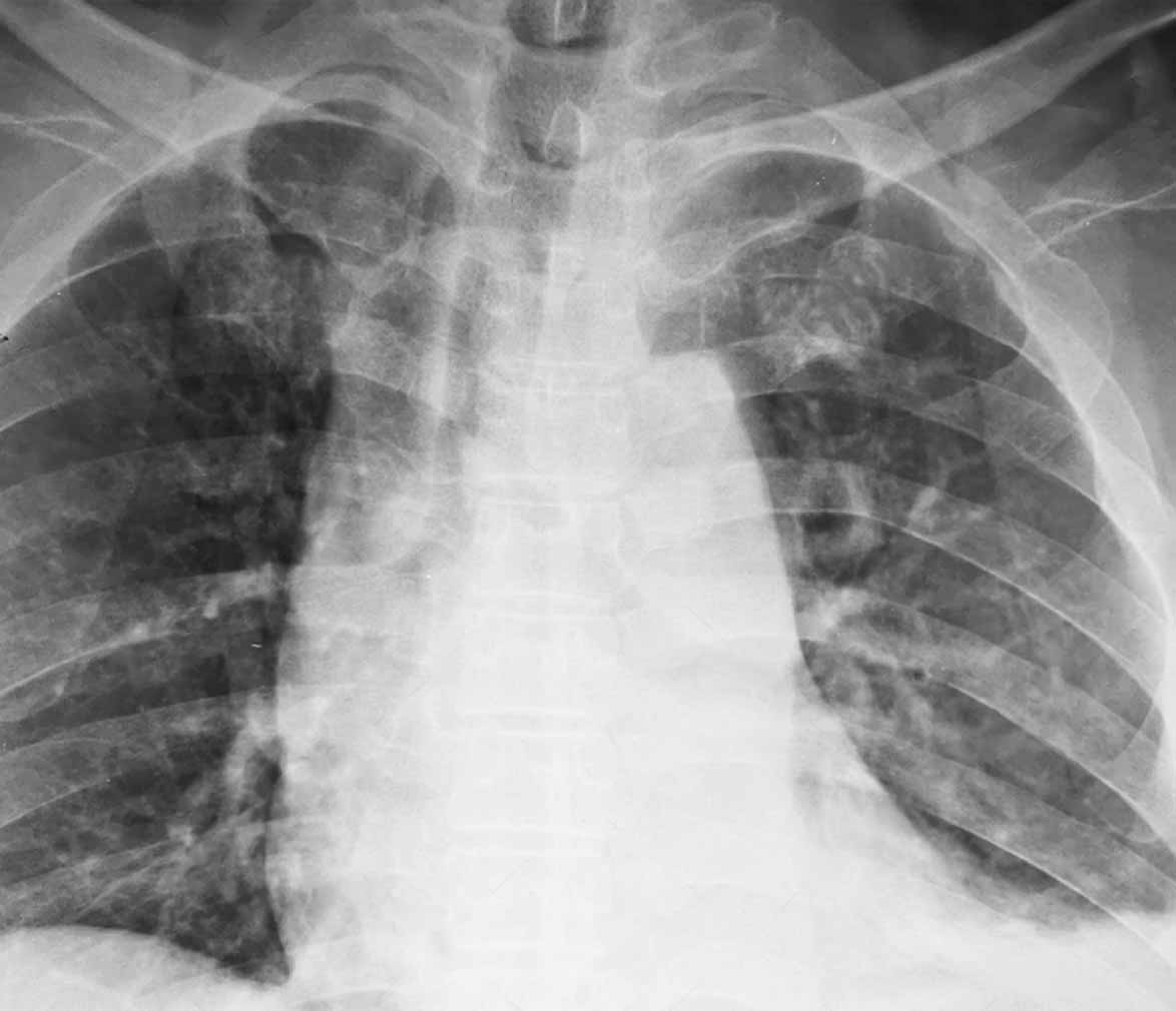
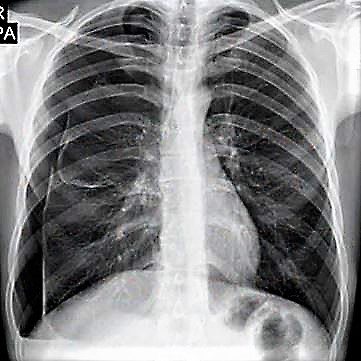
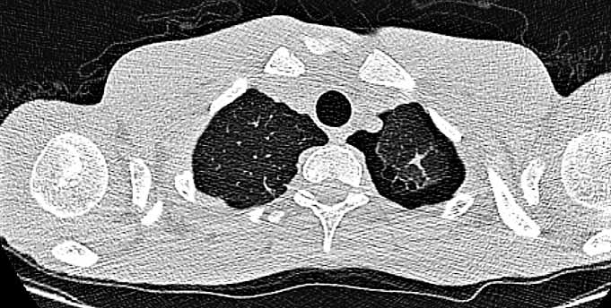
Pneumothorax can be diagnosed by a series of tests and examinations. These include simple chest x-ray and computed tomography. A simple chest x-ray is the most commonly used method to diagnose pneumothorax. Computed tomography is often used to detect the presence of a pneumothorax and to understand its cause.

How is Pneumothorax Treated?
Treatment of a pneumothorax requires medical intervention. Treatment methods include oxygen therapy, tube thoracostomy and surgery (VATS procedure).
A tube thoracostomy is the insertion of a drain through the side chest wall into the pleural cavity. This allows the air accumulated in the lung to be drained and the lung to return to its normal size. In most cases, this procedure is sufficient. However, when this procedure is insufficient, surgery (removal of the diseased area by VATS) is inevitably performed.
VATS is the removal of bubbles in the lung with the help of a camera. The surgical procedure is a treatment method both for therapeutic purposes and to reduce the possibility of recurrence of pneumothorax. Patients can usually be discharged in 2-3 days.
COPD (Emphysema) Surgery
Chronic changes in lung tissue cause permanent damage to the organ over time. Emphysema is one of these diseases. It is closely related to smoking and environmental factors. In emphysema, each inhalation fills the alveoli with air and each exhalation results in a micro level of air trapping, with less of the inhaled air being exhaled. Eventually, the inhaled air is always more than the exhaled air and the lung tissue, which has lost some of its elasticity due to COPD, becomes more swollen than it should be. There comes a point when the person starts to feel short of breath. Various treatments are used to try to slow down and stop the process. However, in progressive cases, if it is thought that other treatments may also fail, appropriate patients may be sent to surgery.
Today, this surgery can be performed with VATS (closed surgery, endoscopic surgery). The swollen lung tissue that is compressing the chest wall and making diaphragmatic movements difficult is removed. Thus, the movements of the lung, which is surrounded by the chest wall and diaphragm, are relaxed and the patient lives a more comfortable respiratory life than before.
Postoperative complications such as air leakage, air trapping in the subcutaneous tissue and bleeding problems are more common than other thoracic surgeries. Therefore, hospitalization periods may be prolonged.
However, at the end of the arduous process, patients’ breathing scores increase and shortness of breath, which was previously experienced even in daily work, decreases. Patients have a better quality of life.
Pneumothorax and Pregnancy
Information about the effects and treatment of pneumothorax during pregnancy. Pneumothorax in pregnancy can pose a serious risk to mother and baby. Treatment may vary depending on the stage of pregnancy and the size of the pneumothorax.
Contact
Address: Ankara Abdurrahman Yurtaslan Oncology Training and Research Hospital Hospital Ankara/TURKIYE
Working Hours:
Weekdays : 08:00 – 17:00
Prof.Dr. Göktürk FINDIK © . All Rights Reserved.
